
Johnson ZM, Yuan Y, Li X, Jashashvili T, Jamieson M, Urata M, Chen Y, Chai Y. 2021. Mesenchymal stem cells and 3D-osteoconductive scaffold regenerate calvarial bone in critical size defects in swine. Stem Cells Translational Medicine. 2021 April 1. doi: 10.1002/sctm.20-0534. PubMed PMID: 33794062.
Category: News
Gamechanging Research in Craniosynostosis by Chai Lab featured in Herman Ostrow School of Dentistry
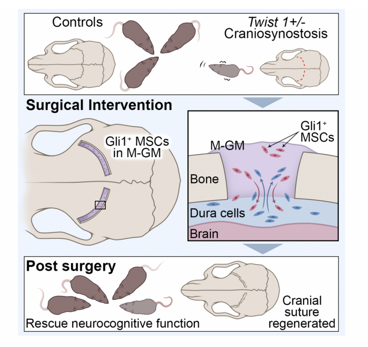
“The discovery could lead to much more than simply fixing an aesthetic condition; it could be life-altering for those experiencing neurocognitive issues resulting from craniosynostosis.” Read full feature here: “Is This The Gamechanger We’ve Been Waiting for in Craniosynostosis?”
New Publication: Cranial Suture Regeneration Mitigates Skull and Neurocognitive Defects in Craniosynostosis
Yu M, Ma L, Yuan Y, Ye X, Montagne A, He J, Ho TV, Wu Y, Zhao Z, Sta Maria N, Jacobs R, Urata M, Wang H, Zlokovic BV, Chen JF, Chai Y. (2021) Cranial suture regeneration mitigates skull and neurocognitive defects in craniosynostosis. Cell. 2021 Jan 7; 184 (1): doi: https://doi.org/10.1016/j.cell.2020.11.037
Publication Highlights
-
-
Twist1 +/− craniosynostosis mice exhibit elevated ICP and cognitive deficits
-
Suture MSCs and resorbable biomaterials can support regeneration of patent suture
-
-
Suture regeneration normalizes Twist1 +/− ICP, skull shape, and cognitive function
-
Suture regeneration offers a biological solution for craniosynostosis patients
New publication: Reciprocal interaction between mesenchymal stem cells and transit amplifying cells regulates tissue homeostasis
Jing J, Feng J, Li J, Zhao H, Ho T, He J, Yuan Y, Guo T, Du J, Urata M, Sharpe P, Chai Y. (2020) Reciprocal interaction between mesenchymal stem cells and transit amplifying cells regulates tissue homeostasis. eLife, Accepted.
New publication: Ror2-mediated non-canonical Wnt signaling regulates Cdc42 and cell proliferation during root development.
Ma Y, Jing J, Feng J, Yuan Y, Wen Q, Han X, He J, Chen S, Ho T, Chai Y. (2020) Ror2-mediated non-canonical Wnt signaling regulates Cdc42 and cell proliferation during root development. Development, Accepted.
New publication: Spatiotemporal cellular movement and fate decisions during first pharyngeal arch morphogenesis
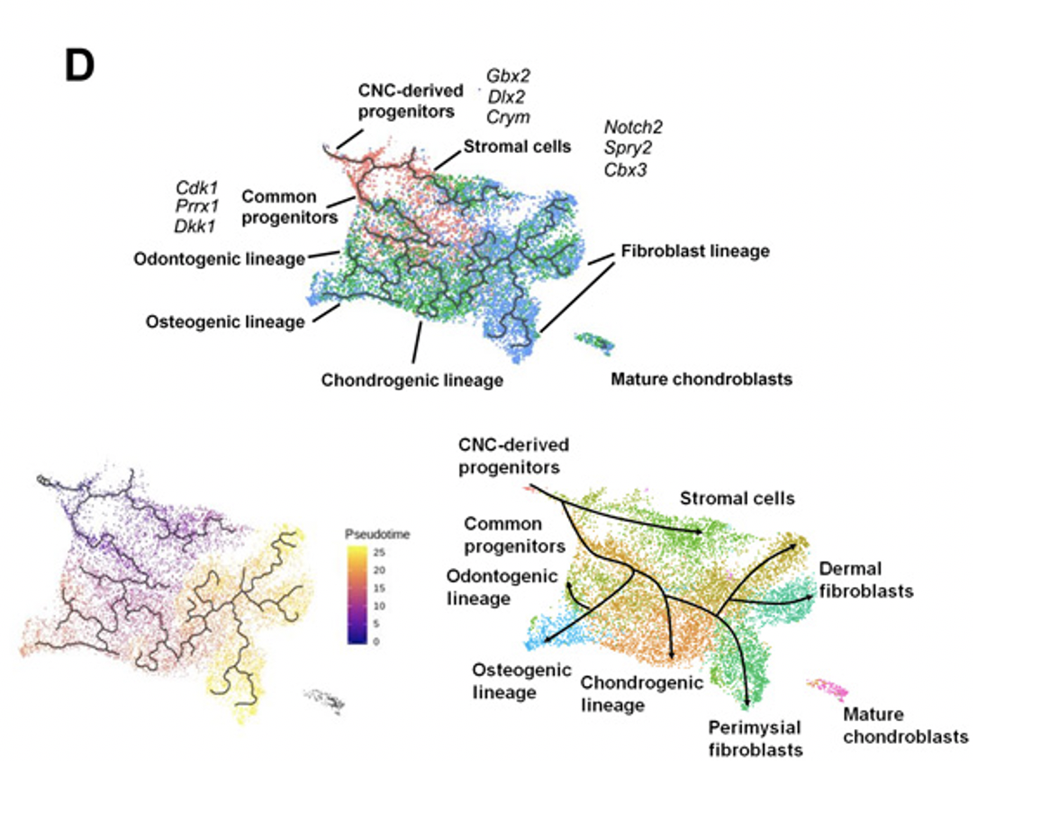
Yuan Y, Loh YE, Han X, Feng J, Ho TV, He J, Jing J, Groff K, Wu A, and Chai Y. (2020) Spatiotemporal cellular movement and fate decisions during first pharyngeal arch morphogenesis. Science Advances. 2020 Dec 16; 6 (51): doi: DOI: 10.1126/sciadv.abb0119
New publication: FaceBase 3: Analytical Tools and FAIR Resources for Craniofacial and Dental Research
Samuels BD, Aho R, Brinkley JF, Bugacov A, Feingold E, Fisher S, Gonzalez-Reiche AS, Hacia JG, Hallgrimsson B, Hansen K, Harris MP, Ho TV, Holmes G, Hooper JE, Jabs EW, Jones KL, Kesselman C, Klein OD, Leslie EJ, Li H, Liao EC, Long H, Lu N, Maas RL, Marazita ML, Mohammed J, Prescott S, Schuler R, Selleri L, Spritz RA, Swigut T, van Bakel H, Visel A, Welsh I, Williams C, Williams TJ, Wysocka J, Yuan Y, Chai Y. (2020) FaceBase 3: Analytical Tools and FAIR Resources for Craniofacial and Dental Research. Development 147: dev191213. doi: 10.1242/dev.191213
New publication: Runx2+ niche cells maintain mesenchymal tissue homeostasis through IGF signaling.
Chen S, Jing J, Yuan Y, Feng J, Han X, Wen Q, Ho TV, Lee C, Chai Y.(2020)
Runx2+ niche cells maintain mesenchymal tissue homeostasis through IGF signaling. Cell Reports, Accepted.
Check out our new lab protocol section
We are beginning to add some tried-and-true lab protocols to our website here. Please check back soon for more.
New publication: Cerebral organoid & mouse models reveal a RAB39b-PI3K-mTOR pathway-dependent dysregulation of cortical development leading to macrocephaly/autism phenotypes
Zhang W, Ma L, Yang M, Shao Q, Xu J, Lu Z, Zhao Z, Chen R, Chai Y, Chen JF. (2020) Cerebral organoid and mouse models reveal a RAB39b-PI3K-mTOR pathway-dependent dysregulation of cortical development leading to macrocephaly/autism phenotypes. Genes Dev. 2020 Feb 27. doi: 10.1101/gad.332494.119. [Epub ahead of print] PubMed PMID: 32115408.